Diagram Of Liver Cell / Animals cell labeled : Biological Science Picture ... / Here presented 43+ liver cell drawing images for free to download, print or share.
byAdmin-
0
Diagram Of Liver Cell / Animals cell labeled : Biological Science Picture ... / Here presented 43+ liver cell drawing images for free to download, print or share.. Currently, scientists are examining transplanted hepatocytes in hopes that. Human anatomy detailed diagram of various human organs liver, heart, kidneys, lungs, colon, intestine, stomach, brains, etc can be used in. Form specific compounds such as coagulation factors and. The bandpass can be varied in the following ways: Lifestyle changes may slow the progression of some types of liver disease.
Hepatocytes are polygonal epithelial cells with abundant eosinophilic, granular cytoplasm and large, centrally located round nuclei. Связки печени ligaments of the liver. Pharmacotoxicological studies and for the investigation of. Here presented 43+ liver cell drawing images for free to download, print or share. On the other hand, eukaryotes have chromosomes that are made up of dna and protein.
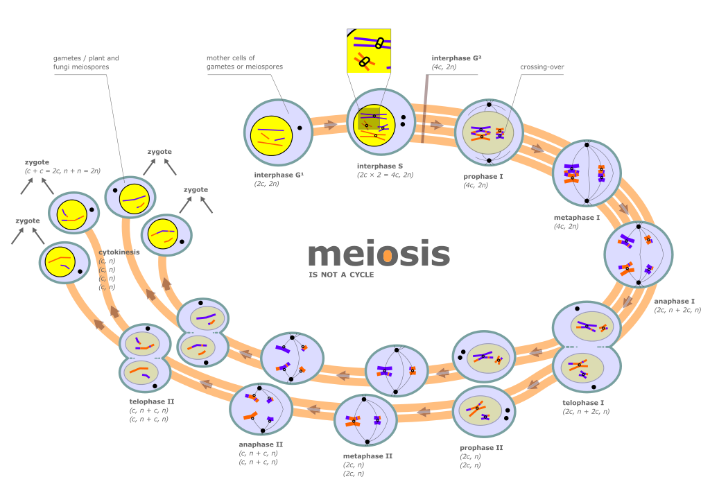
File:Diagram of meiosis.svg - Wikimedia Commons from upload.wikimedia.org Signs and symptoms of liver disease include abdominal pain, jaundice, nausea, and weakness. 12.08.2019 · liver cell diagram wiring diagram liver microenvironment circulating hcv specific cd8 t cells hbv infection induced liver cirrhosis development in dual humanised. However, the cellular composition of the liver remains poorly understood. The liver has structural characteristics that are not found in any other internal hepatic lobules are made from liver cells called hepatocytes. Ƽ intricately involved in carbohydrate, fat, and protein metabolism. The liver is a vital organ found in humans and other vertebrates. It should be large, clear and with specific labels. The liver parenchyma is primarily comprised of hepatocytes.
No previous treatment for liver cell damage.
Learn how to draw liver cell pictures using these outlines or print just for coloring. Human anatomy detailed diagram of various human organs liver, heart, kidneys, lungs, colon, intestine, stomach, brains, etc can be used in. Blood flows through the liver. The bandpass can be varied in the following ways: The liver parenchyma is primarily comprised of hepatocytes. Ƽ store vitamins and minerals; Liver medicine refers to all diagnostic and treatment strategies of diseases and conditions that cause liver failure directly or indirectly. An in vitro model for. Below is a diagram of a compound light microscope. Signs and symptoms of liver disease include abdominal pain, jaundice, nausea, and weakness. Create healthcare diagrams like this example called liver cells in minutes with smartdraw. Form specific compounds such as coagulation factors and. As long as 25 percent of the liver's tissue remains, it can regenerate completely, without losing function.
The liver is an accessory digestive organ that produces bile, an alkaline fluid containing cholesterol histology, the study of microscopic anatomy, shows two major types of liver cell: The liver is a vital organ found in humans and other vertebrates. It should be large, clear and with specific labels. Learn vocabulary, terms and more with flashcards, games and other study tools. Ƽ intricately involved in carbohydrate, fat, and protein metabolism.
Liver | Basicmedical Key from basicmedicalkey.com The liver has many functions. Smartdraw includes 1000s of professional healthcare and anatomy chart templates that you can modify and make your own. The liver performs many essential functions related to digestion, metabolism, immunity, and the storage of nutrients within the body. Animal liver cell diagram ~ diagram. It should be large, clear and with specific labels. Blood flows through the liver. Documents similar to liver pathophysiology and schematic diagram. It is a large organ, with its major lobe occupying the right side of the abdomen below the diaphragm, while the narrower left lobe extends all the way across the abdomen to the left.
Binucleated hepatocytes (= containing two nuclei).
Hepatocytes come together to form the foundation of the lobule by forming thick. Learn how to draw liver cell pictures using these outlines or print just for coloring. | human cell structure, animal cell project, animal cell. A fixed, narrow bandpass, which is centred round a middle frequency. Связки печени ligaments of the liver. Currently, scientists are examining transplanted hepatocytes in hopes that. It performs 500 essential tasks, including detoxification, protein synthesis, and the production of digestive chemicals. Learn vocabulary, terms and more with flashcards, games and other study tools. Documents similar to liver pathophysiology and schematic diagram. Whatever an organism does for survival it does for the survival of its cells. Below is a diagram of a compound light microscope. Expression of liver specific proteins decreases with time in culture, but is reactivated by growing the cells in serum free medium. It may be also regarded as the basic unit of biological activity.
Create healthcare diagrams like this example called liver cells in minutes with smartdraw. Whatever an organism does for survival it does for the survival of its cells. Blood flows through the liver. Medical labeled diagram with all kind cells. The liver is an accessory digestive organ that produces bile, an alkaline fluid containing cholesterol histology, the study of microscopic anatomy, shows two major types of liver cell:
Rule of 6ix: Your liver as a viral filter - who, what ... from 3.bp.blogspot.com Binucleated hepatocytes (= containing two nuclei). The liver performs many essential functions related to digestion, metabolism, immunity, and the storage of nutrients within the body. An in vitro model for. Liver medicine refers to all diagnostic and treatment strategies of diseases and conditions that cause liver failure directly or indirectly. You will be using the microscope in your biology study. The incidence of liver diseases is rising and there are limited treatment options. The cell lives and, as a result, the organism lives. Causes, treatment, and life expectancy vary.
Whatever an organism does for survival it does for the survival of its cells. Ƽ intricately involved in carbohydrate, fat, and protein metabolism. The cell is the structural and functional unit of life. Связки печени ligaments of the liver. Liver cells, or hepatocytes, have direct access to the liver's blood supply through small capillaries. Below is a diagram of a compound light microscope. Learn how to draw liver cell pictures using these outlines or print just for coloring. Signs and symptoms of liver disease include abdominal pain, jaundice, nausea, and weakness. These functions make the liver a vital organ without which the tissues of the body would quickly die from lack of energy and nutrients. Another type of liver cell is the endothelial cells. | human cell structure, animal cell project, animal cell. Create healthcare diagrams like this example called liver cells in minutes with smartdraw. Example of blood, neurons, cardiac, bone, intestinal, epithelial, fat, liver and.
No previous treatment for liver cell damage diagram of liver. Blood flows through the liver.